Neuroscience
Through research in experimental neuroscience and related neuro-theory, we aim to generate novel insights into the nature of mammalian intelligence. Because the neocortex is considered the ‘center’ of mammalian intelligence, we use a combination of neuroimaging, electrophysiology, and data analysis techniques (e.g., miniaturized in vivo caclium imaging) to investigate learning in the neocortex of mice. We train mice on a variety of tasks and then record their neuronal activity in selected cortical brain areas. To understand learning at the single cell level, we record plasticity in individual cortical neurons using patch clamp, calcium, and voltage imaging.
Behavior
The main purpose of the mammalian brain is to generate behavior. The better the brain can generate behavior and the quicker it can adapt to new situations the more intelligent the organism. To better understand biological intelligence, we start our neuroscience investigations by piloting rodent learning paradigms. In a second step, we then combine the new learning paradigm with electro- and opto-physiological methods to record and track neuronal activity in selected areas of the rodent brain.
Miniscope Imaging
We use in vivo miniaturized microscopy to record and track large populations of genetically identified neurons in rodent brains over multiple days and weeks. We record neuronal population activity during active/passive avoidance, fear conditioning, reward/operant conditioning, as well as classical go/no-go tasks.

Single-Cell Patch-Clamp
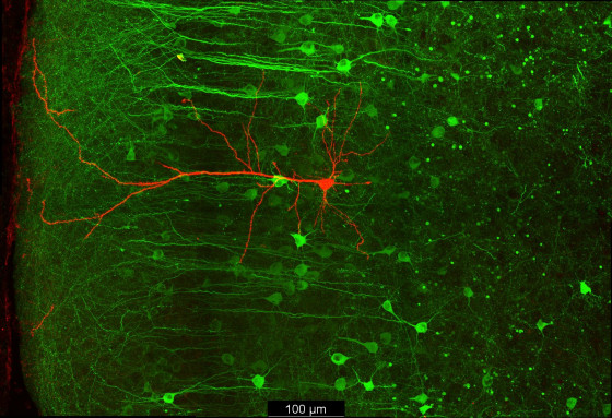
To investigate neural learning at the singe neuron level, we utilize the patch clamp technique in combination with voltage and calcium imaging to monitor cortical L5 and L2/3 pyramidal neurons. In particular, we study how basal and apical input jointly drive the spiking output of pyramidal neurons and how neuronal activity is translated into plasticity. The results out of this line of research will help to inspire new learning rules that we can integrate into the novel, bio-inspired (deep) neuronal networks.
Two-Photon-Imaging
We use in vivo 2P calcium imaging in combination with head fixed learning paradigms in mice to study learning and plasticity in the rodent brain. In contrast to single-photon microscopy, 2P imaging allows to resolve fine dendritic and even synaptic structures and how these are affected by learning. 2P microscopy is a crucial tool to understand processes of learning and plasticity at the single cell, dendritic and synaptic level in awake behaving animals.
